Wednesday, January 6, 2016

BASIC ACCOUNTING CONCEPTS

BASIC  ACCOUNTING  CONCEPTS

     Accounting  is based on certain assumptions.  These assumptions are known as basic  accounting  concepts. Those basic accounting concepts are as follows.


  • Business entity concept : This concept states that the business and its owners are two separate and  distinct  entities. According to this concept, all transactions of the business have to be accounted for from the viewpoint of the business and not from the view point of its owners. The distinction between the business  and its owners is essential in order to ascertain the true picture  of a business.  If the two are not  separated for accounting purposes, the transactions of the busiess will be mixed up with the personal  transactions of its owners  and the true picture of the business can not obtained.


  • Going  concern concept : This concept implies that a business has an indefinite life and it exists for a long period of  time. All business tranactions are performed and recorded from this point of view. The long- term expenditures such as the purchase of land, building and  machinery that the business makes are recorded  in books of  account assuming  that  it will exist and run for a long period  of time.  Their  costs and not the current resale values are recorded spreading over their estimated working  lives. Therefore, the balance sheet always shows fixed assets at cost after subtracting the depreciation.


  • Money measurement concept :  This concept assumes that only those business tranactions which are measured and expressed in monetary terms have to be taken into account. It is so assumed  because money provides a common measure for different goods, services, assets and  liabilities. This concept also assumes that  monetary units such as 'rupee' are stable units in value , but this assumption may not be  true in reality. Therefore, in spite of a decrease in the purchasing power of money, accounting  is performed assuming that the value of money is stable over time.


  • Accounting period concept : The accounting period concept implies that for the purpose of reporting financial information, the whole life of the  business is divided into imaginary  time- intervals. Each time interval is called  an accounting period which is normally  of one year. In Nepal, it begins on the 1st of Shrawn  every year and ends  on the last day of Asad the next year. At the end of each accounting  year, financial statements are drawn to ascertain the  profit  or loss and the financial position of the  business, and are reported  to their users such as owners, managers and creditors.

  • Revenue concept-This concept is also called realization concept. The concept states that revenue is assumed  to be earned when it is realized . According to the concept, revenue is realized when goods  are transferred to the buyers  and services are provided to the clients for  cash, or for assets or in  anticipation of  realizing the value of sales on a future date. It is not  necessary that the revenue must be realized in cash.  Besides , revenue is  earned in the period when it is realized. However, revenue  realized is always net  of goods returned from the customer  and bad debts.


  • Cost concept :  This concept implies that the cost of anything  such as a service or an asset is recognised when it is incurred and not when cash  is paid for it.  According to the concept, the  cost is assumed to be incurred when the service or the asset is used to generate  revenue .  Besides, the concept assumes that  the asset is taken into account at the cost of its purchase and not at its market value. This concept , however does not mean that the cost of purchase appears in the books every year. Since an asset  has a limited life, its cost  is written  off every year over its life. Thus, the books show the asset at the purchasing  cost less its depreciation up- to- date.


  • Matching concept: This concept provides guidelines as to how the profit or loss of a business should be determined. The  concept , therefore, states that  revenue earned in a period has to be matched with  the expenses incurred  in the same period so as to find  out the true  profit or loss  of the business. While matching the expenses with the revenue , the latter should be realized first and then only the expenses  relating to the revenue should be recognized . Any expense or revenue of the previous  or the next year  should not be matched with those of this  year. If they are matched, the true profit or loss can not be  ascertained.


Epigenetics and Cancer


Epigenetics and  Cancer


INTRODUCTION
  
        
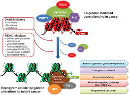
         Epigenetics  is a rapidly evolving study of molecular genetics and biological research. The advancement in the understanding of different  biological activity like DNA methylation, chromatin structure, transcriptional  activity and histone modification has resulted in the development  of epigenetics. Epigenetics changes influence gene transcription  without alteration in the DNA  sequence. The term '' epigenetics was first used by Conrad  Waddington in 1939 to describe ' the causal interaction between the genes and their products, which bring  the phenotype into being.'' In  present  era the term  epigenetics has broadened to include heritable and  transient / reversible changes in gene expression that is not accompanied  by a change in the DNA  sequence. There are two type  of major of major  epigenetic  modification: those occurring  at the DNA level (DNA methylation) and  those occurring at the chromatin  level ( chromatin  remodeling). DNA  Methylation is an  enzyme driven chemical change to the DNA  sequence that most commonly occurs at CpG dinucleotides.  Chromatin remodeling  occurs via modification of the histone residues  by enzymes  primarily  on  the N- terminal  tails and ultimately effects  the interaction  of DNA  with chromatin modifying  proteins. Both DNA methylation and histone modification are associated with silencing critical  tumor suppressor genes and activating oncogones involved in cancer.


       Most  of the traditional  molecular studies on cancer are on identifying the genetic mutation or on tumor suppressor gene. Recently, more studies are now focused in discovering new biomarkers  that are epigenetically silenced in early carcinogenesis.  It  is also seen that almost half of the  tumor suppressor genes that  causes familial  cancer through mutations can  also get inactivated with  promoter hypermethylation  in sporadic  cancer. Increasing  evidence suggests  that epigenetic changes  play a key role in  cancer development.

          
          The various  genetic  markers has been used for the early  tumor detection, prognostic prediction and explaining  the genetic pathway of carcinogenesis. But the epigenetics marker has  gained popularity in recent year particularly the promoter hypermethylation which has various  advantages over  a genetic marker.  First promoter hypermethylation is  much more common than genetic alteration  in cancer.  Second promoter hypermethylation occur in the same defined region of that gene  in  all  form of cancer in comparison to wide  range of mutational  variations occur within a specific  gene. Thus epigenetic detection of promoter                                                                                             hypermethylation  will be both efficient and 
                                                                       cost- effective method of tumor detection.   


DNA METHYLATION AND EPIGENETIC GENE SILENCING

DNA  METHYLATION AND EPIGENETIC GENE SILENCING


     DNA methylation is a reversible chemical modification of the cytosine in the CpG islands  of promoter sequences, catalyzed by a family of DNA methyltransferases. DNA  methylation does not change  the genetic information but it just alters the readability of the DNA  and results  in the  inactivation of gene by  subsequent transcript repression.  CpG island  are the regions  in DNA  that  contains  many adjacent cytosine and guanine nucleotides. The ''p '' in CpG  refers to the phosphodiester bond between  the cytosine   and  the guanine. These islands  occur  in  approximately  40% of the promoters  of human  genes. These 
islands occur  in approximately 40%  of the promoters  of human genes. DNA methylation  plays  a critical  role in the control  of cellular  process  including  embryonic development , transcription, X- chromosome inactivation and  genomic imprinting. DNA  methylation occurs in the C5 positions of cytosines that precedes guanines and  are called dinucleotide CpGs. The CpG dinucleotides are not found frequently throughout the human genome and present 20 % of its  expected frequency. But approximately half of the human gene promoter regions have CpG rich areas of 0.5 to 2 kb in length. In which CpG dinucleotide frequency are higher than expected . These CpG rich areas are often known  as CpG islands. . The majority (94%) of CpG island  remain  unmethylated  in normal cell. But particular subgroups of promoters CpG are  methylated  such as tissue  and germ line specific genes. In general , CpG  island methylation causes gene silencing. The  methylated CpG  island  also recruit   histone deacetylases  and other factor involved  in
transcriptional silencing .  In activation  of tumor suppressor genes through  hypermethylation of CpG islands within promoters regions is a major event in carcinogenesis. Hypermethylation of CpG
  islands within promoter regions  is a major  event in  carcinogenesis . Hypermethylation of CpG  island also has silencing effect on miRNA  in cancer. Micro RNA are short , 18-22  nucleotide, noncoding RNas that regulate many cellular functions including cell proliferation, apoptosis and differentiation by silencing specific target  genes through translational repression or mrNa degradation.

Tuesday, December 1, 2015

Laparoscopic


          Laparoscopic   Management   OF   Large   Hiatus   Hernia

INTRODUCTION


   The Management of large hiatal hernias is difficult and their operative repair can be technically challenging . The concept of a large  hiatal  hernia, however, has not been clearly defined. They have been classified by various  authors according to whether the hiatal defect  is larger or  smaller  than 5 cm and or / or their  contents. Aly et al considered hiatal hernias to be large when more than 50 percent  of the stomach has migrated into the chest.  Andujar  defined  it as the presence  of more than one- third of the stomach in the thoracic cavity. Carlson et al in his study has  defined  hiatal  hernias to be large if the  hiatal defect is larger than 8 cm which was later  modified to 5-6 cm. We consider paraesophageal hernias to be large  when more than half of the stomach has migrated into the chest or if the hiatal defect is larger than 5 cm in size.
    
    Hiatal hernias are classified into type I to IV depending on the position of the gastroesophageal junction in relation to the diaphragmatic  hiatus. According to the conventional classification, type III and type IV can be considered to be large hernias. Type III hernias, which is a mixed sliding and paraesophageal hernia (PEH)  occurs predominantly in the elderly population . The end stage of a hiatus hernia  is an intrathoracic stomach in which the whole stomach  migrates into the chest by rotating 180 degrees along its longitudinal axis with the caedia and the pylorus as fixed points  and includes other organs including colon, omentum, small bowel , liver and spleen (TypeIV).
   
     Surgical correction of large hiatal hernias is indicated because of unsatisfactory outcomes after long- term medical  management and potentially disastrous consequences of gastric incarceration or volvulus  in large paraesophageal hernias. Fundamental steps involved in large hiatal hernia repair include a tension free reduction of the esophagus  and the stomach into the abdomen with complete excision of the hernial sac, reapproximation  of the hiatus, and subdiaphragmatic  fixation of the stomach, with many authors advocating the additional of an antireflux procedure.


     Recurrence rates after laparoscopic surgery is a controversial and unresolved  issue. A few early studies have shown alarmingly  high recurrence rates. Some possible patient related and procedure  related mechanisms are  in appropriate postoperative activity  of the patient immediately after surgery, inadequate excision of the sac, inadequate mobilization of the esophagus, inadequate crural closure secondary to widely spaced crura sutured  under  tension , or a postoperative rupture of crurorraphy due to continuous excursion of the diaphragm.
   
     After complete reduction of all the hernial contents, the necessity of complete excision  of the  hernial  sac cannot be understated. IN  earlier series recurrence rates of up to 20 percent have been  reported after inadequate excision  of the hernial  sac . Although  sac removal is tedious and difficult,it is one of the most  crucial  steps of large paraesophageal  hernia repair. If  circumferential  reduction of the sac  is not done and a portion  of the  sac of new  hernia formation has been  practically  left behind. Fluid collections in the unresected sac leading to postoprative dysphagia have also been reported in literature.
   
     A number  of methods have been adopted to reduce the risk of postoperative recurrences and include the use of Teflon pledgets to prevent crural sutures from cutting through, complete detachment of the sac from the hiatus and mediastinum, complete excision of the sac, adequate mobilization of the esophagus and use of mesh cruroplasty in patients with a large hiatus hernia to achieve a tension free hiatal.


SURGICAL PRINCIPLES


    Although the need for surgical repair is undebated, controversies exist concerning the best surgical  approach  whether open or laparoscopic, the presence of short esophagus and the need for an esophageal lengthening procedure, crurorraphy or a tension free mesh repair, subdiaphragmatic  fixation of the stomach , the need for an antireflux procedure, whether total or partial fundoplication and the indication for prosthetic  reinforcement of the hiatus.

GALLSTONES

                                           GALLSTONES


      Biliary tract  disease is the second  most common  non- obstetric  surgical problem, though it affects only 1 in 1600  to 10000 pregnancies. Cholelithiasis has been documented in 10  percent  of pregnancies and  cholecystitis reportedly affects  0.1 percent  of pregnant patients.
        Pregnancy related  physiological  changes : a. Progesterone causes smooth  muscle relaxation  and  a decrease  in gallbladder tone.  Weakened  contractions and  decreased  emptying  lead to increased  gallbladder  volume  during  fasting  and after eating. In  turn, biliary  stasis  contributes  to cholesterol  crystal sequestration ,  theoretically leading to the  formation  of sludge and stones. b. Elevated  estrogen levels during  pregnancy may further  increase the lithogenicity  of bile. c. Lower  gallbladder  ejection  fractions  and increasing  parity seem  to increase the risk of  sludge formation. A high pre-pregnancy body mass  index  also  may increase the  risk  of sludge  formation. Despite these  physiologic changes, it is unclear if  pregnancy increases  the incidence of gallstones  and  cholecystitis . In  a German  population  study looking  at 1111 females, current pregnancy and the number of prior pregnancies were not associated with an increased  risk.

       The clinical presentation of acute  cholecystitis is similar  to the non- pregnant patient. The pregnant - patient who has right upper  quadrant  tenderness  should undergo ultrasound  evaluation  first because it is  noninvasive and quickly obtained.  MR cholangiography can be suspected  but not demonstrated on  ultrasound.  Symptomatic  cholelithiasis often is managed initially with  a conservative approach,  delaying elective cholecystectomy until after delivery. If conservative  management fails, or if repeated hospitalizations are required, especially  in the same  trimester, cholecystectomy is indicated. Recent studies have  shown, earlier surgical intervention  for biliary tract disease in pregnancy is safe with reduced hospital stay, reduced use of  medications, lower rates  life- threatening complications and lower preterm deliveries. The  laparoscopic cholecystectomy  has been performed safely in all trimesters. The use of an  open  technique for entry, insufflations to 12 mm Hg, and  maintaining a left lateral decubitus  position minimize risk to the fetus and  help maintain adequate placental blood flow during  surgery.
        Patients presenting with acute cholecystitis and symptomatic choledocholithiasis  during pregnancy  should be considered  in a higher  risk  category. If  complications such  as cholangitis or gallstone  pancreatitis develop, maternal mortality  approaches 15 percent, and fetal loss occurs in 60 percent of cases . 


        Surgical approaches  include open cholecystectomy with choledochotomy  or laparoscopic  cholecystectomy with ERCP (endoscopic retrograde cholangiopancreatography ) with sphincterotomy  for  stone extraction or stent placement has been shown  to be  safe during pregnancy.  Although  not routinely recommended, intraoperative  cholangiography   is safe  after fetal organogenesis is complete  and does not increase the risk   of preterm  labor or adverse fetal outcomes.

APPENDIX

                                                      APPENDIX


     Appendicitis is the most common non- obstetric surgical complication and the most common gastrointestinal disorder  requiring  surgery during pregnancy. It accounts  for 25 percent of surgeries for non- obstetric  indications in  pregnancy and complicates every  1 in 1500 to 2000 pregnancies. The incidence of perforated  appendicitis in pregnant women is 43 percent compared to 4-9 percent in the non- obstetric  population.  This increased  incidence may be due to delay in diagnosis and reluctance to operate in pregnancy. Maternal and fetal morbidity and  mortality correlate with perforation and  its associated complications. Uncomplicated appendicitis has a 3-5 percent  fetal loss rate with negligible maternal mortality. Appendix perforation, however, is associated with a 20-35 percent fetal loss rate and 4 percent maternal mortality. Appendix  perforation , however , is associated  with  a 20-35 percent  fetal  loss rate and 4 percent  maternal  mortality. Maternal  mortality rates have  dropped  significantly in the  recent  years with prompt surgical  intervention , newer antibiotics and  surgical  techniques. 

      The preterm contractions  caused  by  uterine irritation from perforation peritonitis result in preterm delivery in 5-14 percent. This incidence is similar between  open  and laparoscopy. In the  first trimester the  appendix remains in its  normal anatomic  position. The appendix  undergoes progressive displacement cephalad  and laterally with advancing pregnancy. After 24 weeks gestation, the  appendix is shifted superiorly  above the right iliac crest , and the tip of the appendix is rotated  medially toward the uterus. By  late pregnancy, the appendix may be closer to the gallbladder than MCBurney's point, occupying the right upper quadrant . This change may alter the location of the pain, making diagnosis difficult. As the  peritoneum is displaced  from the appendix  and cecum by the growing uterus, the increased separation of  the visceral  and parietal peritoneum  decreases  the somatic  sensation of pain and compromises the ability to  localize pain  on examination. The enlarging uterus interfere with the ability  of the omentum and bowel to wall off the inflamed appendix.  Diffuse peritonitis from perforation is  facilitated by this inability of the omentum to isolate the infection. The appendix  returns  to its normal  position by the tenth postpartum day. However, modern clinical experience does not confirm this assertion, with recent studies demonstrating  that the most frequent  location of pain  remains in the right lower quadrant, regardless of trimester. 
               

     Symptoms of appendicitis often  are confused with normal  pregnancy related conditions, particularly in the third trimester. Pain in the right lower quadrant is the most  common and reliable symptom  of appendicitis along with the usual features of appendicitis. Rectal  and pelvic tenderness may not be present when the appendix  is displaced by the uterine enlargement. Leukkocytosis is normal in pregnancy  and so not a good  indicator. Ultrasound Scan is useful  in the first  and second trimester. MRI is safe in pregnancy . CT  Scan should be reserved for cases where Ultrasound and MRI are non- diagnostic. Despite reluctance to operate on a pregnant patient, immediate surgical  intervention is indicated when a  diagnosis of appendicitis is made. The choice of surgical procedure is  based on uterine size and experience  of the  surgeon.  The only indication  of delay  is active labor, and in these  cases the surgery is performed immediate  postpartum.
Add caption


      Diagnostic and operative  laparoscopy  is reasonable  before 20 weeks gestation  and is as safe as open surgery. When  performing  laparoscopy  open entry(Hassan)  is preferable to avoid inadvertent veress or trocar  entry into uterus. Trocar placement  needs to be changed according  to the  size of the uterus. Beyond the late second trimester, laparoscopy  becomes more technically challenging . Appendicitis is confirmed in 36-50  percent of cases. Accuracy of diagnosis  in the  first trimester is greater.  A higher false positive rate is acceptable  in pregnant women, because  any delay in diagnosis may compromise maternal and fetal  well - being .

Tuesday, November 3, 2015

THE DIABETIC FOOT


    An  important  underlying  cause leading to diabetic foot problem is neuropathy. Sensory  neuropathy  leads  to a loss of  protective  sensation. Foot  trauma  is unrecognised and leads to ulceration. The ulceration is often the portal of entry for bacteria, leading to cellulites and/ or abscess formation. Motor neuropathy can lead to asymmetric  muscle atrophy, foot deformity (equines deformity) and altered  biomechanics. This  leads to areas of high pressure during standing or walking and repeated  trauma  that may go unrecognised  because of sensory deficit. Autonomic neuropathy results in loss of sweating and dry skin that leads to cracks and  fissures and a portal of entry for bacteria. Diabetes is also associated with an increased risk of peripheral arterial disease and it can be a major factor in non- healing of foot ulcerations.
 
     Diabetic patients can have significant foot infection, with much less pain and no pronounced systemic inflammatory response. A high index of suspicion is therefore required to diagnose foot infection in patients with diabetes.
                                                   

     The skin of the foot is a highly specialized organ. The plantar skin consists of a complex array of fascia, fibrous septae and tangential shearing forces that occur during walking. The dorsal skin is bound to the underlying extensor retinaculum.  Infection tracks along fascial  planes and tendon sheaths. The location of a diabetic foot wound will usually lead surgeon to the underlying cause. An  ulcer at the posterior border of the heel is usually the result of chronic pressure from prolonged contact with bedding, friction from rubbing against rough bed sheets and lack of elevation. An ulcer about the plantar foot is almost always due to i) excessive pressure and time between the foot and the contact surface, ii) neuropathy and iii) deformity of the foot ( equines contracture). Understanding  the underlying cause will allow an effective wound care. The single best means of reducing pressure on the sole of the foot  is to employ non- weight bearing of the involved  limb through use of crutches or walker. This  may not always be practical but effort should be made to emphasize compliance. Use of standard off- loading shoes are also recommended.
   
    Pain in a neuropathic  foot is usually related to an underlying infection. After a thorough surgical preparation, in the emergency room, to remove debris and allow proper evaluation, the wound is probed to determine its depth and tissues involved. Osteomyelitis should be considered if the wound is deeper than the dermis layer. Most patients with diabetes who present  with a severe foot infection have chronically poor glycemic  control, chronic anaemia, poor nutrition and deficient clinical care. Therefore, laboratory studies and necessary management is essential  before embarking on active treatment.


    An important initial  step in treating limb threatening diabetic foot infection is to perform a timely and adequate surgical  debridement. This entails surgical excision of all nonviable and/ or infected  tissue. The plantar spaces are opened by longitudinal incisions with division  of plantar fascia. When pus is present in flexor tendon sheaths, these are opened and drained. In order to appropriately evaluate the viability of the soft tissues and the underlying structures, surgical  debridement should be performed without the use of a tourniquet. If there is exposed bone or suspicion of osteomyelitis, cultures are obtained of this tissue. Wound is irrigated and meticulous hemostasis achieved. Most diabetic foot infections are treated with an empirically selected antibiotic regimen until cultures and sensitivity are available . In a limb threatening infection or osteomyelitis, intravenous therapy should be initiated and followed if possible by oral agents. The use of topical antibiotics has a limited place as may produce the development of resistant strains of colonized surface bacteria.

    A patient with diabetes may not give the typical history of claudication because of associated  neuropathy or lack of activity. It is therefore  important to evaluate the limb for peripheral arterial disease even  in the absence of symptoms. If pedal pulses are not clearly palpable, further vascular studies are indicated. An ankle- brachial index (ABI) should be obtained.

      There is a close association between peripheral arterial disease and coronary disease making then both a high risk for a traditional open bypass procedure. The endovascular options include percutaneous angioplasty  with or without a stent . These procedures cab be performed under local  anaesthesia and sedation and with a high rate of limb salvage .

      The standard treatment of diabetic foot ulcer includes adequate off- loading of weight, frequent ulcer debridement , wound care, treatment of infection and revascularization of ischemic limb. This standard  care in many controlled trials  has resulted in healing of a number of foot ulcers.  However, newer therapeutic  modalities that can improve healing also need to be explored.

     Despite recent advances in surgical and radiologic vascular techniques, a fair number of patients with critical limb ischemia are not eligible for a revascularization procedure.  This is because of anatomic location of the lesion, the extent of the disease or extensive co-morbidity . No  effective pharmacologic  therapy is available  and amputation is often  the only option left.  The cost of managing a patient after amputation has been estimated to be almost  twice that of a successful limb salvage . Therefore, exploring new strategies for ischemic limbs is of major importance.  Bone marrow derived progenitor cells have been identified as a potential new therapeutic target.

    Normal wound healing is a intricate process involving various  cell types, coordinated processes, and complex signaling  interactions.  In a diabetic wound , many of these  responses to inflammatory mediators, matrix production, angiogenesis, and wound contraction have all been  poor and contribute to delayed  healing of a diabetic wound.

     Cell - based therapy is an attractive approach for the treatment of wounds with multiple impairments. Mesenchymal stromal  cells (MSCs) are the multipotent cells derived from stroma  of bone marrow  and other tissues. The local delievery  of  MSC to a diabetic wound might  correct wound healing impairment both indirectly by reversing local  growth - factor deficiency, and directly by improving wound contraction through  interaction with the extracellular  matrix. The decrease in wound size  might  have the potential to offset diabetic - related wound - healing impairment  significantly.
                                                 

       In a  randomized  controlled  trial in 28 diabetic patients with critical limb ischemia, Huang et al reported improvement in limb ischemia and foot ulcers.  By far the most studies of cell therapy have used intramuscular implantation method or intraarterial  injection. Progenitor cell- based  therapy may have great clinical potential.